
Introduction to Radiological Assessment Of Fractures
Accurate radiological assessment of fractures is essential for proper fracture diagnosis and treatment planning. This guide covers the key imaging modalities—from basic radiographs to advanced MRI—and explains how to optimize their use in clinical practice.
The Golden Rule: Radiograph Fundamentals
High-quality radiographs are non-negotiable for proper fracture evaluation. Follow the “Rule of Twos” for optimal imaging:
1. Two Orthogonal Views
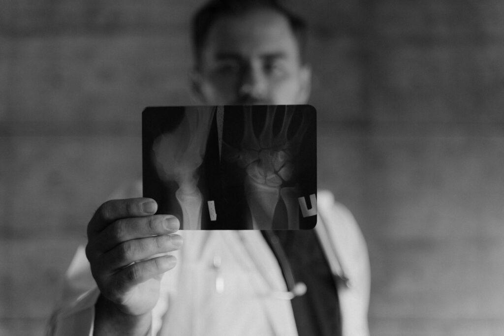
- Always obtain AP (anteroposterior) and lateral views (90° apart)
- Example: A wrist fracture requires both views to assess displacement
2. Two Joints
- For diaphyseal fractures, image the joints above and below
- Why? Reveals rotational malalignment and hidden injuries
- Exception: Isolated joint injuries (e.g., distal radius) may only need local views
3. Two Time Points
- Some fractures (e.g., scaphoid) appear only after 10-14 days (due to bone resorption)
- Clinical tip: Repeat imaging if initial X-rays are negative but symptoms persist
4. Two Limbs (When Needed)
- Compare with the asymptomatic side for:
- Congenital anomalies
- Pediatric physeal line confusion (Use an atlas first!)
Specialized Radiographic Techniques
Traction Films
- Taken while applying traction to clarify complex fracture patterns
- Often requires sedation/anesthesia
Stress Views
- Assess joint stability (e.g., suspected ligament tears)
- Common uses: Ankle syndesmosis injuries
Anatomy-Specific Series
- Some fractures need special views:
- Scaphoid: Ulnar deviation + scaphoid view
- Calcaneus: Harris axial view
- Shoulder: Y-view or Velpeau axillary
Advanced Imaging Modalities
Ultrasound (POCUS)
- Best for soft tissues:
- Achilles/quadriceps tendon tears
- Rotator cuff injuries
- Detects small joint effusions
CT Scans
- 3D reconstructions for complex fractures (Fig. 1.26)
- Key uses:
- Intra-articular fractures (e.g., tibial plateau)
- Spinal trauma
- Preoperative planning
MRI
- Gold standard for:
- Occult fractures (scaphoid, femoral neck)
- Soft tissue damage (ligaments, menisci)
- Bone marrow edema (stress fractures)
Ancillary Diagnostic Tools
Joint Aspiration
- Indications:
- Acute joint pain + swelling
- Suspected septic arthritis or gout
Blood Tests
- Essential for:
- Infection markers (CRP, ESR)
- Malignancy workup (Ca²⁺, ALP)
- Pre-op assessment (Hb, electrolytes)
Clinical Pearls: Avoiding Pitfalls
| Scenario | Solution |
|---|---|
| Negative X-rays but high clinical suspicion? | Repeat films in 10-14 days or get MRI |
| Pediatric physeal confusion? | Reference an atlas before extra X-rays |
| Complex intra-articular fracture? | CT scan for surgical planning |
| Suspected ligament injury? | Stress views or MRI |
Conclusion of Radiological Assessment Of Fractures
- Start with proper radiographs (follow the Rule of Twos)
- Use advanced imaging judiciously:
- CT for complex bony anatomy
- MRI for soft tissue/occult fractures
- Don’t ignore clinical correlation—imaging is just one piece of the puzzle
Pro Tip: For scaphoid fractures, MRI early avoids delayed diagnosis complications.
Read more ortho topics: Orthopedics Surgery
Read further from authentic sources:
- American College of Radiology (ACR) Appropriateness Criteria
https://www.acr.org/Clinical-Resources/ACR-Appropriateness-Criteria - Radiological Society of North America (RSNA) – Fracture Detection
https://pubs.rsna.org/doi/full/10.1148/rg.2020190079 - NIH – Radiation Safety in Medical Imaging
https://www.ncbi.nlm.nih.gov/books/NBK565909/ - Journal of Orthopaedic Trauma – Imaging Recommendations
https://journals.lww.com/jorthotrauma/pages/default.aspx - AAOS – Occult Fracture Diagnosis
https://orthoinfo.aaos.org/en/diseases–conditions/fractures-broken-bones/ - International Emergency Radiology Society
https://www.emergencyradiology.org/