Introduction to Comprehensive Overview of Liver Anatomy
The liver, the largest internal organ in the human body, is a vital hub for metabolism, detoxification, and digestion, making its anatomy a cornerstone of medical knowledge. This guide provides a detailed exploration of the liver’s structure, covering its ligaments, dual blood supply (from the hepatic artery and portal vein), segmental anatomy as defined by Couinaud’s eight segments, and venous drainage through the hepatic veins into the inferior vena cava. Additionally, it delves into the functional units of the liver, such as the hepatic lobules, which facilitate its metabolic and detoxification processes. Understanding these anatomical details is essential for diagnosing liver conditions, performing surgical resections, and advancing medical research, making this overview an invaluable resource for medical students, surgeons, and healthcare professionals alike.
1. Liver Anatomy Overview
- Size and Location:
The liver is the largest organ in the body, weighing approximately 1500 grams. It is located in the right upper abdominal cavity, beneath the diaphragm, and is protected by the rib cage. It has a reddish-brown color and is encased in a fibrous sheath called Glisson’s capsule. - Ligaments and Peritoneal Reflections:
The liver is anchored in place by several ligaments formed by peritoneal reflections:- Falciform Ligament:
- A remnant of the umbilical vein, it runs from the umbilicus to the liver between the right and left lobes.
- Divides the left lateral and left medial segments along the umbilical fissure.
- Anchors the liver to the anterior abdominal wall.
- Left Triangular Ligament:
- Located on the superior surface of the left lobe.
- Dividing its anterior and posterior folds allows mobilization of the left lobe from the diaphragm and exposure of the left lateral wall of the IVC.
- Right Triangular Ligament:
- Fixes the right lobe to the undersurface of the right hemidiaphragm.
- Division of this ligament allows mobilization of the liver from under the diaphragm.
- Coronary Ligaments:
- Extend from the triangular ligaments and anchor the liver to the diaphragm and retroperitoneum.
- Lesser Omentum:
- A thin peritoneal reflection between the stomach and liver.
- Contains the hilar structures (hepatic artery, portal vein, and bile duct) in its free edge.
- Falciform Ligament:
2. Segmental Liver Anatomy
The liver is divided into functional segments based on vascular and biliary anatomy:
- Cantlie’s Line:
- The functional plane dividing the liver into right and left lobes.
- Extends from the gallbladder fossa to the inferior vena cava (IVC).
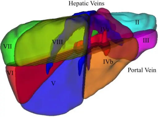
- Couinaud’s Segmentation:
- The liver is divided into eight functional segments, each with its own arterial, venous, and biliary inflow and outflow.
- Segments I–VIII:
- Segment I: Caudate lobe.
- Segments II and III: Left lateral segment.
- Segment IV: Left medial segment (subdivided into IVA and IVB).
- Segments V–VIII: Right lobe (V and VIII form the right anterior lobe; VI and VII form the right posterior lobe).
- Brisbane Terminology:
- Standardizes liver anatomy and resection terminology:
- First-Order Division: Left and right hemilivers.
- Second-Order Division: Sections within each hemiliver.
- Standardizes liver anatomy and resection terminology:
3. Blood Supply (Liver Anatomy)
The liver has a dual blood supply, with 75-80% from the portal vein and 20-25% from the hepatic artery.
- Hepatic Artery:
- Arises from the celiac trunk, which branches into the common hepatic artery.
- The common hepatic artery divides into the gastroduodenal artery and hepatic artery proper, which further branches into the right and left hepatic arteries.
- Variations:
- Replaced Right Hepatic Artery: Present in 10-20% of cases, arising from the superior mesenteric artery (SMA).
- Replaced Left Hepatic Artery: Present in 10-15% of cases, arising from the left gastric artery.
- Portal Vein:
- Formed by the confluence of the splenic vein and superior mesenteric vein.
- Divides into right and left portal veins at the hilum.
- Left Portal Vein: Supplies segments II, III, and IV.
- Right Portal Vein: Supplies segments V–VIII.
4. Structures in the Hilum of the Liver
The porta hepatis is the hilum of the liver, containing the following structures within the hepatoduodenal ligament:
- Hepatic Artery:
- Lies above and medial to the bile duct.
- Branches into the right and left hepatic arteries, with the right hepatic artery crossing the bile duct either anteriorly or posteriorly.
- Portal Vein:
- Lies posteriorly within the ligament.
- Divides into right and left branches at the hilum.
- Bile Duct:
- Lies anterior and lateral within the ligament.
- The common hepatic duct is joined by the cystic duct to form the common bile duct (CBD).
- The left hepatic duct has a longer extrahepatic course (~2 cm).
5. Venous Drainage
The liver drains blood via the hepatic veins into the IVC:
- Hepatic Veins:
- Right Hepatic Vein: Drains segments V–VIII.
- Middle Hepatic Vein: Drains segment IV and parts of V and VIII.
- Left Hepatic Vein: Drains segments II and III.
- The middle and left hepatic veins usually join within the liver parenchyma before entering the IVC.
- Inferior Hepatic Veins:
- Short vessels that drain directly into the IVC.
- Retrohepatic IVC:
- Lies in a groove on the posterior liver surface.
- The right adrenal vein drains into the IVC at this level.
6. Functional Units: Hepatic Lobules
The functional units within the liver segments are the hepatic lobules:
- Structure:
- Composed of plates of liver cells (hepatocytes) separated by hepatic sinusoids.
- Sinusoids carry blood from the portal tracts (containing branches of the hepatic artery and portal vein) to the central vein (a tributary of the hepatic vein).
- Function:
- Blood flows through the sinusoids, enabling liver functions such as bile formation.
- Bile flows in the opposite direction to blood, draining via bile duct tributaries in the portal tracts.
7. Clinical Considerations
- Liver Resection:
- Understanding segmental anatomy and vascular variations is critical for safe liver resections.
- Resections are based on anatomical lines to preserve maximal functioning liver.
- Liver Regeneration:
- The liver can fully regenerate after partial resection.
- Surgical Mobilization:
- Division of ligaments (e.g., falciform, triangular) allows full mobilization of the liver during surgery.
Summary of Key Points
- Ligaments:
- Falciform, triangular, coronary, and lesser omentum anchor the liver and contain critical structures.
- Segmental Anatomy:
- The liver is divided into eight segments based on Couinaud’s classification, with Cantlie’s line separating the right and left lobes.
- Blood Supply:
- Dual supply: 75-80% from the portal vein and 20-25% from the hepatic artery.
- Variations in arterial anatomy (e.g., replaced hepatic arteries) are common.
- Venous Drainage:
- Three hepatic veins drain into the IVC, with the middle and left hepatic veins often joining before entry.
- Functional Units:
- Hepatic lobules are the functional units, enabling metabolic and biliary functions.
Also read: