Introduction
The surgical management of acute and chronic wounds is a cornerstone of effective wound care, requiring precise techniques to ensure optimal healing, functional recovery, and minimal scarring. Acute wounds, such as traumatic injuries, bites, and high-pressure injection injuries, demand immediate debridement, repair, and closure, while chronic wounds, including those caused by hidradenitis suppurativa or necrotizing soft tissue infections, often require advanced surgical interventions like skin grafts, flaps, and negative pressure wound therapy. This guide delves into the detailed surgical approaches for managing both acute and chronic wounds, emphasizing evidence-based practices to achieve the best outcomes.
1.1. Principles of Acute Wound Management
- Examination:
- Assess the wound site, structures involved, and mechanism of injury.
- Evaluate movement, sensation, and pain.
- Follow ATLS (Acute Trauma Life Support) principles to avoid missing injuries (e.g., stab wounds in the back).
- Debridement:
- Remove contaminated and devitalized tissue.
- Excise until bleeding tissue is encountered, except for nerves, vessels, and tendons.
- Use copious saline irrigation or pulsed jet lavage.
- Avoid blind clamping of vessels to prevent nerve damage.
- Muscle viability is judged by color, bleeding pattern, and contractility.
- Repair:
- Primary repair of structures (e.g., bone, tendon, nerve, vessel) in tidy wounds.
- Nerve repair: Use 8/0 or 10/0 monofilament nylon under magnification (loupes or microscope).
- Vessel repair: Use similar techniques for arteries like the radial or ulnar artery.
- Tendon repair: Early active mobilization to minimize adhesions between the tendon and tendon sheath.
- In contaminated or untidy wounds, perform multiple debridements before definitive repair (e.g., second-look surgery).
- Primary repair of structures (e.g., bone, tendon, nerve, vessel) in tidy wounds.
- Skin Closure:
- Ensure closure without tension to allow for edema during the inflammatory phase.
- Use flaps or grafts for coverage if needed.
- Flaps: Bring in a new blood supply to cover exposed structures (e.g., tendon, bone, nerve).
- Grafts: Depend on the recipient site for nutrition. Meshed split-thickness grafts can be used for larger wounds.
1.2. Specific Acute Wounds
- Bites:
- Cleanse thoroughly and explore for damage.
- Treat with antibiotics for anaerobic and aerobic organisms.
- Human bites: Common on the ear, nose, and metacarpophalangeal joint (e.g., boxing-type injuries).
- Puncture Wounds:
- Explore to the limit of tissue blood staining.
- X-ray to rule out retained foreign bodies.
- Needlestick injuries: Follow protocols for hepatitis and HIV risks.
- Degloving Injuries:
- Open or closed injuries where skin and subcutaneous fat are stripped from underlying fascia.
- Excise nonviable skin until punctate dermal bleeding is observed.
- Use split-thickness skin grafts harvested from degloved skin.
- Fluoroscein can be used intravenously to map viable skin under ultraviolet light.
- Compartment Syndromes:
- Diagnose based on severe pain, pain on passive movement, sensory disturbance, and elevated compartment pressures (>30 mmHg).
- Treat with fasciotomy via longitudinal incisions to decompress affected compartments.
- Avoid late fasciotomy in crush injuries to prevent myoglobinuria and renal failure.
- High-Pressure Injection Injuries:
- Wide surgical exposure and debridement of toxic substances.
- Preoperative X-rays to detect air or lead-based paints.
- High amputation rates (>45%) if not treated promptly.
2. Surgical Management of Hidradenitis Suppurativa (HS)
2.1. Operative Approaches
- Incision and Drainage:
- For abscesses during disease flares.
- High risk of recurrence.
- Technique:
- Palpate abscess and identify area of fluctuance.
- Administer local anesthetic.
- Make incision in the thinnest epithelial coverage.
- Break up loculations and irrigate with sterile saline.
- Leave the wound open and pack with dressing or loosely close over a Penrose drain.
- CO2 Laser Excision:
- For Hurley Stage 2 or 3 disease.
- Excises hair follicles to reduce recurrence.
- Deroofing or Wide Local Excision:
- For severely scarred or tunneling disease.
- Comparable recurrence rates (~25%).
- Wound management options:
- Primary closure.
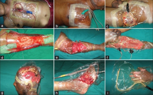
- Negative pressure wound therapy.
- Split-thickness skin grafting.
- Local flaps (e.g., Limberg flap for axillary HS).
3. Surgical Management of Necrotizing Soft Tissue Infections (NSTIs)
3.1. Principles of Surgical Treatment
- Early Aggressive Debridement:
- Critical for survival.
- Remove all necrotic tissue, including skin, subcutaneous fat, and fascia.
- Intraoperative tissue cultures to guide antibiotics.
- Reassessment:
- Return to the operating room within 24 hours for further debridement if infection progresses.
- Wound Management:
- Initial management with topical antimicrobial gauze (e.g., Dakin’s solution).
- Transition to negative pressure wound therapy or delayed primary closure once infection is controlled.
3.2. Surgical Technique
- Fasciotomy:
- For compartment syndromes associated with NSTIs.
- Longitudinal incisions to decompress affected compartments.
- Avoid late fasciotomy in crush injuries to prevent myoglobinuria and renal failure.
4. Surgical Management of Chronic Wounds
4.1. Principles of Surgical Intervention
- Debridement:
- Remove necrotic tissue and biofilms.
- Convert chronic wounds into acute wounds to restart the healing process.
- Biopsy:
- Rule out malignant conversion (e.g., Marjolin’s ulcer).
- Skin Grafts and Flaps:
- For large defects or exposed structures.
- Split-thickness skin grafts for superficial wounds.
- Flaps for deeper wounds with exposed bone, tendon, or nerve.
4.2. Specific Techniques
- Negative Pressure Wound Therapy (NPWT):
- Promotes granulation tissue formation.
- Reduces wound size and prepares for closure.
- Delayed Primary Closure:
- After infection control and adequate debridement.
- Local Flaps:
- For coverage of defects in specific regions (e.g., Limberg flap for axillary HS).
5. Surgical Focus on Wound Closure and Healing
5.1. Primary Intention
- Wound edges are opposed surgically.
- Minimal inflammation and scarring.
- Ideal for clean, surgical incisions.
5.2. Secondary Intention
- Wound is left open.
- Heals by granulation, contraction, and epithelialization.
- Increased inflammation and scarring.
- Used for contaminated or infected wounds.
5.3. Tertiary Intention (Delayed Primary Intention)
- Wound is initially left open due to contamination.
- Edges are later opposed when conditions are favorable.
- Results in a less satisfactory scar compared to primary intention.
6. Key Surgical Techniques
- Debridement:
- Essential for contaminated, infected, or necrotic wounds.
- Use saline irrigation, pulsed jet lavage, or sharp excision.
- Skin Grafts:
- Split-thickness grafts for superficial wounds.
- Full-thickness grafts for better cosmetic outcomes.
- Flaps:
- Local flaps for small defects.
- Regional or free flaps for larger defects or exposed structures.
- Negative Pressure Wound Therapy:
- Enhances granulation tissue formation.
- Prepares wounds for closure or grafting.
7. Specific Surgical Procedures
7.1. Tendon Repair
- Early active mobilization to minimize adhesions between the tendon and tendon sheath.
- Use of 8/0 or 10/0 monofilament nylon for repair under magnification.
7.2. Vessel Repair
- Repair of vessels like the radial or ulnar artery using microsurgical techniques.
7.3. Nerve Repair
- Use of 8/0 or 10/0 monofilament nylon under magnification (loupes or microscope).
7.4. Skin Flaps
- Limberg Flap: Used for axillary HS after wide local excision.
- Local Flaps: For coverage of defects in specific regions.
7.5. Split-Thickness Skin Grafts
- Harvested from degloved nonviable skin and meshed to cover raw areas after debridement.


